
Your body builds plasmalogens throughout early life—but it does not keep building them forever. Research reveals a distinct rise-and-fall pattern in these specialized ether phospholipids, with consequences that touch every organ from the brain to the heart. This guide maps what the peer-reviewed literature tells us about when plasmalogens peak, why production falters, and what downstream effects ripple through cellular health as levels drop.
What This Guide Covers
- What Makes Plasmalogens Different From Other Phospholipids
- The Rise Phase: Birth Through Mid-Adulthood
- The Decline Phase: Mid-Life and Beyond
- The Peroxisome Connection: Where Biosynthesis Breaks Down
- Tissue-Specific Patterns of Loss
- Downstream Consequences of Falling Plasmalogen Levels
- How Plasmalogen Levels Are Measured
- Key Takeaways
- Frequently Asked Questions
What Makes Plasmalogens Different From Other Phospholipids
Plasmalogens belong to a specialized subclass of glycerophospholipids distinguished by a vinyl ether bond at the sn-1 position of the glycerol backbone. This bond is not merely a structural footnote—it gives plasmalogens unique antioxidant capacity and membrane-modulating properties that conventional diacyl phospholipids lack.
The two major classes found in mammalian tissues are ethanolamine plasmalogens (PlsEtn) and choline plasmalogens (PlsCho). PlsEtn species are particularly enriched in the brain and nervous system, where they support membrane fluidity, vesicle fusion, ion transport, and synaptic signaling. Their biosynthesis begins inside peroxisomes and finishes in the endoplasmic reticulum—a two-organelle pathway with no redundant backup.
The Rise Phase: Birth Through Mid-Adulthood
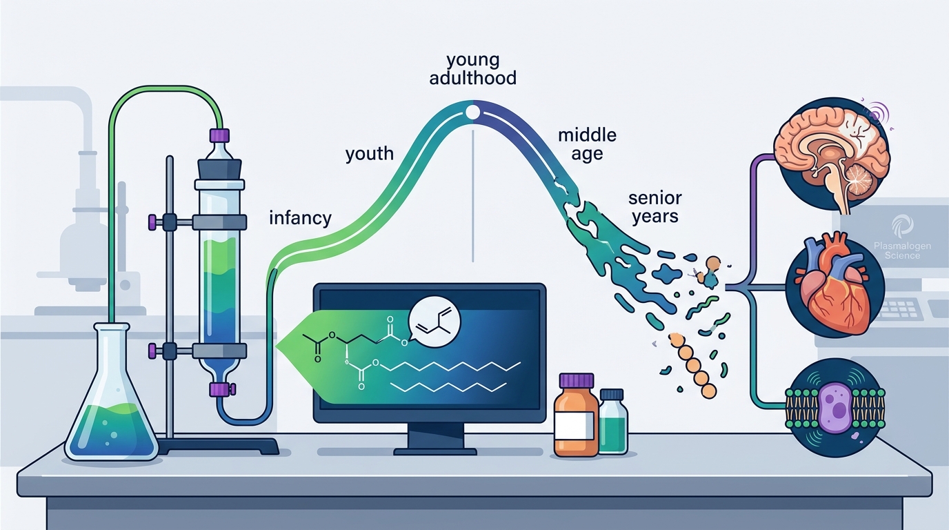
Plasmalogen accumulation is an active process during development. From infancy through the third and fourth decades of life, the body steadily manufactures and incorporates plasmalogens into cell membranes across virtually every tissue. Epidemiological data demonstrate that brain plasmalogen levels increase linearly up to approximately 30–40 years of age, reflecting the maturation and peak performance of the peroxisomal biosynthetic machinery.
During this rise phase, plasmalogens contribute to robust myelination, efficient synaptic communication, and strong antioxidant buffering capacity within neuronal membranes. The heart, kidneys, and immune cells also accumulate substantial plasmalogen reserves during these years.
The Decline Phase: Mid-Life and Beyond
After their peak in early-to-mid adulthood, plasmalogen levels begin a measurable decline. Research published in Frontiers in Physiology shows that plasmalogen levels keep increasing linearly up to 30–40 years of age and then, by 70 years of age, a significant linear decrease is observed (Rouser and Yamamoto, 1968). Particularly after age 50, the drop becomes clinically meaningful, as the brain becomes more vulnerable to oxidative stress and inflammation.
This is not simply a gradual tapering. In individuals with neurodegenerative conditions, the decline is accelerated well beyond normal aging trajectories. Studies measuring serum PlsEtn in over 400 clinically demented subjects found circulating levels significantly lower than in non-demented age-matched controls, and the degree of depletion tracked with dementia severity.
What Accelerates the Decline
Several factors push plasmalogen levels lower faster than chronological aging alone would predict:
- Chronic inflammation — persistent inflammatory signaling consumes plasmalogens as they sacrifice their vinyl ether bond to neutralize reactive oxygen species.
- Metabolic stress — insulin resistance, obesity, and type 2 diabetes are all associated with lower plasmalogen scores. One large population study found a significant inverse association between a composite plasmalogen score and the prevalence and incidence of type 2 diabetes.
- Environmental oxidative burden — exposure to pollutants, smoking, and excessive alcohol consumption generates reactive species that degrade plasmalogens faster than they can be replenished.
The Peroxisome Connection: Where Biosynthesis Breaks Down
Plasmalogen biosynthesis depends entirely on functional peroxisomes. The initial enzymatic steps—catalyzed by glyceronephosphate O-acyltransferase (GNPAT) and alkylglycerone-phosphate synthase (AGPS)—take place exclusively within the peroxisomal matrix. No alternative cellular pathway exists to produce the alkyl-ether precursors that define plasmalogens.
This makes peroxisomal health a rate-limiting variable for plasmalogen supply. Aging is accompanied by progressive declines in key peroxisomal functions, including catalase activity, fatty acid β-oxidation, and plasmalogen biosynthesis, resulting in increased oxidative stress, lipid dysregulation, and alterations in membrane composition. Research in human B cells confirms an age-related decline in peroxisome biogenesis, with decreased expression of PEX19 and reduced import of peroxisomal matrix enzymes in aged cells.
The enzyme dihydroxyacetone-phosphate acyl-transferase (DHAP-AT), which is the rate-limiting enzyme in plasmalogen biosynthesis, shows reduced activity when peroxisomal protein import is compromised. This creates a bottleneck: even if raw substrates are available, the cellular factory that assembles plasmalogen precursors runs at diminished capacity.
The Peroxisome–Mitochondria Axis
Peroxisomal dysfunction does not stay contained within peroxisomes. Falling plasmalogen output disrupts mitochondrial fission dynamics, as recent research in Experimental Cell Research (2026) demonstrated using Drosophila models. When peroxisomal plasmalogen biosynthesis declined with age, stress-induced mitochondrial fission was impaired—compromising the cell’s ability to clear damaged mitochondria and maintain energy production.
This peroxisome–mitochondria crosstalk means that plasmalogen depletion simultaneously undermines both the membrane integrity side (plasmalogens in cell membranes) and the energy production side (mitochondrial quality control) of cellular resilience.
Tissue-Specific Patterns of Loss
Plasmalogen depletion does not occur uniformly across the body. The tissues most enriched in plasmalogens during youth are often the most vulnerable to age-related loss.
Brain and Nervous System
Plasmalogens are found in especially high levels in neuronal membranes, where they support synaptic vesicle formation, neurotransmitter release, and myelin sheath maintenance. Post-mortem analyses show that reduced cognition in the elderly is associated with low levels of plasmalogens and high levels of lipid rafts, amyloid plaques, and neurofibrillary tangles in the temporal cortex. In Alzheimer’s disease specifically, brain PlsEtn levels are reportedly lower than in age-matched controls.
Heart
Cardiac tissue is another major plasmalogen reservoir. PlsEtn and PlsCho species in the heart contribute to membrane fluidity in cardiomyocytes, and their reduction with age may partially explain increasing cardiac stiffness and arrhythmia risk in older adults.
Immune Cells
Plasmalogens play roles in phagocytosis by macrophages. Research in aged rodent brains shows that microglia become less active in phagocytosis compared to those in young brains, and this functional decline may be linked to reduced brain plasmalogen levels.
Downstream Consequences of Falling Plasmalogen Levels
The effects of plasmalogen depletion cascade through multiple layers of cellular function:
1. Compromised Antioxidant Defense
The vinyl ether bond at sn-1 acts as a sacrificial scavenger of reactive oxygen species. As plasmalogen concentrations drop, membranes lose this frontline defense, making remaining lipids, membrane proteins, and even DNA more vulnerable to oxidative damage.
2. Reduced Membrane Fluidity and Signaling
As levels decrease, cell membranes become less flexible and more vulnerable to oxidative damage. This shift in membrane biophysics impairs receptor function, ion channel behavior, and the formation of lipid rafts essential for signal transduction.
3. Synaptic and Cognitive Decline
In the brain, reduced plasmalogen availability may affect how well neurons communicate and how effectively the brain maintains its structure. Mouse studies demonstrate that plasmalogen supplementation can enhance synaptic plasticity in the hippocampus of aged animals, with increased expression of synaptophysin—a key marker of functional synapses.
4. Neuroinflammation
Aging brains are often accompanied by increased neuroinflammation and synaptic loss, which may be attributable to aging-dependent microglial dysfunction. Plasmalogen depletion contributes to this cycle by impairing microglial phagocytic capacity and promoting pro-inflammatory signaling.
5. Metabolic Vulnerability
Population-level data link lower plasmalogen scores with increased prevalence and incidence of type 2 diabetes, suggesting that plasmalogen depletion is both a marker and a potential contributor to broader metabolic dysregulation.
How Plasmalogen Levels Are Measured
Clinically, plasmalogen status is most commonly assessed through serum or plasma lipidomics using liquid chromatography–tandem mass spectrometry (LC-MS/MS). Key analytes include specific PlsEtn species, particularly those carrying DHA (22:6) at the sn-2 position, which are the most sensitive to age-related depletion.
Researchers have developed composite metrics such as the Plasmalogen Biosynthesis Value (PBV)—a combination of three key PlsEtn species—and the Plasmalogen Score (Pls Score), which can serve as an independent modifiable marker of metabolic health. These scoring systems allow clinicians and researchers to categorize individuals as plasmalogen-sufficient or plasmalogen-deficient relative to age-adjusted reference ranges.
Post-mortem brain tissue analysis provides the most direct measure of tissue-level plasmalogens but is naturally limited to research contexts. Serum levels have been shown to correlate with brain levels, making blood-based testing a practical proxy for assessing central nervous system plasmalogen status.
Key Takeaways
- Plasmalogen levels rise steadily from birth through approximately age 30–40, then decline significantly by age 70.
- The decline accelerates in the presence of chronic inflammation, metabolic disease, and neurodegenerative conditions.
- Peroxisomes are the sole site of plasmalogen precursor synthesis, and age-related peroxisomal dysfunction is a primary driver of declining plasmalogen production.
- Falling plasmalogen levels compromise antioxidant defense, membrane fluidity, synaptic integrity, mitochondrial dynamics, and immune function.
- Serum lipidomics via LC-MS/MS can quantify plasmalogen status, with composite scores offering a clinically useful biomarker of metabolic and neurological health.
- The brain, heart, and immune cells are among the most vulnerable tissues when plasmalogen supply diminishes.
Frequently Asked Questions
At what age do plasmalogen levels start to decline?
Research shows that plasmalogen levels in the brain increase until approximately 30–40 years of age, after which a significant decline begins. The decrease becomes more pronounced around age 50 and continues through later decades. Chronic inflammation or metabolic stress can trigger earlier or more rapid depletion.
Why do plasmalogen levels decrease with age?
The primary cause is declining peroxisomal function. Peroxisomes are the only organelles capable of initiating plasmalogen biosynthesis, and their protein import machinery and enzymatic activity deteriorate with age. Additionally, increased oxidative stress in aging tissues consumes existing plasmalogens faster than they can be replaced.
Can you measure your plasmalogen levels?
Yes. Plasmalogen levels can be measured in blood serum using liquid chromatography–tandem mass spectrometry (LC-MS/MS). Researchers have developed composite scores, such as the Plasmalogen Biosynthesis Value, that combine multiple PlsEtn species into a single clinically interpretable metric. These serum measurements correlate with brain plasmalogen levels.
Is plasmalogen decline linked to Alzheimer’s disease?
Multiple studies have found that serum and brain PlsEtn levels are significantly decreased in Alzheimer’s disease patients compared to age-matched controls. The degree of plasmalogen depletion correlates with dementia severity, and patients with normal plasmalogen levels showed no cognitive decline over a one-year follow-up period, suggesting a protective association.
Do plasmalogens affect mitochondrial function?
Yes. Plasmalogen biosynthesis and mitochondrial dynamics are linked through peroxisome–mitochondria crosstalk. When peroxisomal plasmalogen production declines, it impairs stress-induced mitochondrial fission—a quality-control process essential for removing damaged mitochondria. This connection means plasmalogen depletion can compromise cellular energy production alongside membrane integrity.
This article is provided by Plasmalogen Science for educational purposes. It is not intended as medical advice. Consult a qualified healthcare provider before making decisions based on this content.